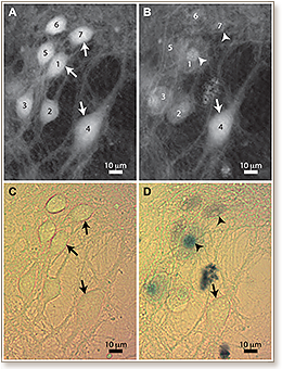
 Digital holography provides a non-invasive measurement of the quantitative phase shifts induced by cells in culture, which can be related to cell volume changes. It has been shown previously that regulation of cell volume, in particular as it relates to ionic homeostasis, is crucially involved in the activation/inactivation of the cell death processes. We thus present here an application of digital holographic microscopy (DHM) dedicated to early and label-free detection of cell death. Methods and Findings: We provide quantitative measurements of phase signal obtained on mouse cortical neurons, and caused by early neuronal cell volume regulation triggered by excitotoxic concentrations of L-glutamate. We show that the efficiency of this early regulation of cell volume detected by DHM, is correlated with the occurrence of subsequent neuronal death assessed with the widely accepted trypan blue method for detection of cell viability. Conclusions: The determination of the phase signal by DHM provides a simple and rapid optical method for the early detection of cell death.